

Overview
There are two types of cartilage in the knee: articular cartilage and meniscus. Articular cartilage is a smooth cartilage that covers the bone surfaces within the knee joint and helps with protection and reducing friction of the joint during movement.
Each knee has two meniscus. These are semi-circles of fibrocartilage that act as shock absorbers in the knee.
Cartilage contains no blood vessels and so does not heal well on its own. Therefore there are surgical techniques to repair, regenerate and replace this cartilage.
These procedures are more appropriate for people who have specific, small and well locslised cartilage defects rather than wide spread joint arthritis.
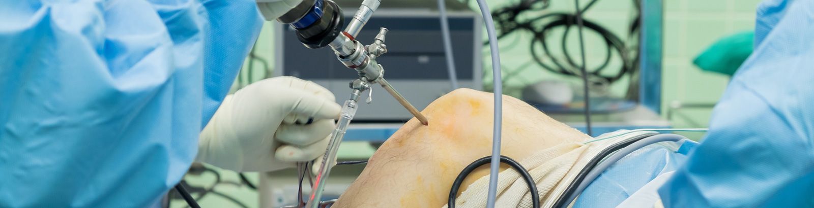
Surgery is usually performed using arthroscopy – a type of keyhole surgery using small cuts and specialised instruments.
The main procedures are:
- Chondroplasty or Cartilage Repair – also called abrasion chondroplasty. The joint is washed out to remove any loose tissue and the edges of damaged or frayed cartilage are trimmed to make them smooth. Damaged cartilage may be repaired at the same time. This can be done for articular cartilage and menisci.
- Microfracture – also called bone marrow stimulation. Tiny holes are made in the bone beneath the damaged cartilage releasing blood and bone marrow into it. Stem cells then stimulate the production of new cartilage. This is only done for articular cartilage.
- Cartilage replacement – also called mosaicplasty. Small plugs of healthy cartilage from non-weight bearing areas of the joint, such as the side of the knee, are removed and used to replace small areas of damaged cartilage. This is only done for articular cartilage
For those where these procedures are ineffective or if joint damage is particularly severe, replacing the joint with an artificial one may be necessary. Your surgeon will go through the options with you.
The operations
- This operation is usually done under general anaesthetic
- This operation can take up to an hour
- Two or three small incisions are made at the front of your knee for the camera (arthroscope) and other instruments to perform the procedure
- Salty water (saline) is passed through the knee to make visualisation of the knee structures easier. This fluid is drained at the end of the procedure
- The procedure is carried out using specialised arthroscopic instruments
- Local anaesthetic and painkillers are injected into the knee to minimise discomfort after surgery
- Stitches are used to close the wounds and a tight bandage is applied
After the operation
- Pain relief – good pain relief is important and you will be given this during your procedure. Some people need more than others so please ask for help if you are in any discomfort following your procedure. You can expect some pain that may last up to a few weeks
- Hospital stay – most patients usually leave the hospital on the same day
- Physical activities – Partial weight bearing is crucial for early recovery and will be part of your rehabilitation programme
- Showering and bathing – you need to keep your wound well covered using the waterproof dressing provided whilst showering. When bathing do not submerse wound in water as dressing are not designed to withstand this. This should be done for at least the first 10 days
- Swelling and bruising – Ice: applied for 10-30 minutes at a time. Wrap ice in a plastic bag or towel to prevent ice-burn. Elevation: reduces swelling. Prop up leg using a pillow when sitting or lying in bed. Aim to keep above the level of the heart. Swelling and bruising may take up to 8 weeks to completely disappear.
Your surgeon and physiotherapist will discuss with you issues surrounding:
- Returning to work
- Using your knee following your operation
- Driving
- Anticipated length of rehabilitation
Possible complications
All surgery has associated risks. These risks are higher in some patients than others. Risks include:
- Anaesthetic – your anaesthetist will discuss risks associated with the anaesthetic with you.
- Infection – there is a small risk of wound infection following your operation that is minimised by giving you antibiotics. If at any time you notice a fever or increased pain, swelling or redness around you wound, please contact us urgently.
- Nerve or blood vessel damage – this can sometimes occur after surgery and if severe may require a return to theatre.
- Deep vein thrombosis – this is a blood clot in the leg. You may be given blood thinning medication and support stocking to help prevent this. If you notice a pain in your calf then please contact us urgently.
- Failure to improve – symptoms may not be cured by the surgery and in a small proportion of cases the discomfort may be worse than prior to surgery.